Saturday, January 23, 2021


The viruses that prey on human diseases
Share on Linkedin
Though it has fallen out of favour in the West, phages have been in use for more than a century (Credit: Romain Lafabregue/AFP/Getty Images)
Once scorned as Soviet pseudoscience, phage therapy is gaining ground as a potential solution to antibiotic resistance but regulatory challenges may be its biggest hurdle.

Article continues below

T

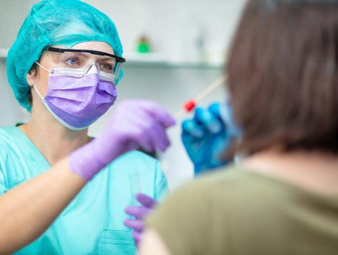
Three years ago, Esteban Diaz was advised by his doctors to get on the lung transplant list after a life-long battle with cystic fibrosis. The disease causes excessive production of mucus in the lungs and pancreas, leaving patients extremely vulnerable to bacterial infections. In the 47-year-old Frenchman’s case, the antibiotics he had been prescribed since childhood were no longer effective against incessant infections caused by Pseudomonas aergonisa, a bacteria now classified as a superbug.

Instead, Diaz (not his real name) travelled to Georgia, a former Soviet state on the Black Sea, to undergo phage therapy, a medical treatment he says cleared up his infections within days and relieved him of the persistent fatigue, relentless coughing and breathlessness that plagued him for decades.

Phages or bacteriophages are viruses that naturally prey on bacteria by infecting and replicating within them until they burst out, killing their microbial host. There are billions of phages on Earth, and they have co-evolved with the bacteria they prey on for millennia, helping to keep their numbers in check.

Their therapeutic use was first pioneered in 1919 by Felix d’Herelle, a French-Canadian microbiologist who used phages to cure a boy suffering from severe dysentery. However, the discovery of penicillin in 1928 and its subsequent commercial production by the 1940s unleashed the antibiotic era, effectively supplanting phage therapy.

You might also like:

The therapeutic role of phages might have been all but forgotten if not for the collaboration between d’Herelle and George Eliava, a young Georgian scientist who had travelled to France in 1923. He had arrived with the aim of studying the development of vaccines but instead turned his attention to phages after meeting d’Herelle at the Pasteur Institute.

Eliava returned to Georgia and invited d’Herelle to help set up the world’s first research institute and therapeutic centre dedicated to bacteriophages, just as the country was being absorbed into the Soviet Union.

Sadly, like thousands of intellectuals of the time, Eliava fell foul of Josef Stalin’s regime and was executed in 1937. But Soviet patronage of the research and development of therapeutic phages continued at the institute Eliava founded, years after the Western world sidelined the approach.

Story continues below

After the fall of the Soviet Union, the Eliava Institute continued research into the disease-fighting abilities of bacteriopages (Credit: Vano Shlamov/AFP/Getty Images)

After the fall of the Soviet Union, the Eliava Institute continued research into the disease-fighting abilities of bacteriopages (Credit: Vano Shlamov/AFP/Getty Images)

“Phage therapy was part of the standard health care system during the Soviet Union,” says Mzia Kutateladze, director of the Eliava Institute. “Depending on the health status of the patient and the type of infection, doctors would make a call on whether to use phages or antibiotics or a combination of both.”

The institute, however, faced severe difficulties in the years following the break-up of the Soviet Union. Some researchers resorted to storing phage cultures in their own homes to save them. But it would soon play a key role in reintroducing the world to the scope and potential of phage therapy.

“It took a long time before people were convinced that phages can be used therapeutically," says Kutateladze. "But antibiotic resistance supported the necessity to find alternatives.” The institute faced enormous challenges when it started presenting its work internationally in the late 1990s. But in 2001, it received its first foreign patient soon after a conference in Montreal – a Canadian suffering from a bacterial bone infection called osteomyelitis that antibiotics hadn’t been able to cure. The treatment worked, and thanks to a flurry of news articles which followed, international patients started trickling into the Eliava Institute.

By the fourth day of treatment it was as if someone had taken back my sickness – Esteban Diaz

The World Health Organization (WHO) has called antimicrobial resistance (AMR) a global health crisis, with up to 30 million people expected to be affected by 2050. For cystic fibrosis patients like Diaz, antibiotic resistance was the inevitable fallout of a lifetime of prescription to the drugs.

“From the time I was seven years old till I was 17… every three months, I would be systematically bombarded with two different kinds of antibiotics – this was the protocol back in the day,” says Diaz. By the time he was in his 30s, he had also developed chronic tinnitus as a side effect of the continued use of aminoglycosides, the most common antibiotic family used to treat pseudomonas infections like his. By his 40s, resistance had set in and double lung transplantation was the only option his doctors in France could suggest to prolong his life.

After coming across a documentary about the Eliava Institute’s phage therapy on a French TV channel, he booked a trip. “By the fourth day of treatment it was as if someone had taken back my sickness. I slept through the night for the first time in years. It’s hard to describe… I could almost sense the oxygen coursing through my lungs. It was incredible,” he says.

Since his first visit, Diaz regularly returned to Tbilisi to stock up on oral doses of phages preparations that have helped keep subsequent infections in check. That was until he ran out of phages in March this year, just as Georgia closed its borders in its effort to tackle the spread of the coronavirus. As soon as travel restrictions were loosened, Diaz returned for another round of treatment that he said immediately eased a persistent cough he had picked up in the meantime.

The institute has been a world leader in bacteriophage research since the 1930s (Credit: Pearly Jacob)

The institute has been a world leader in bacteriophage research since the 1930s (Credit: Pearly Jacob)

But his treatment hasn't been without complications. Diaz fears he might lose his benefits if he is found to have travelled to Georgia for treatment, especially during a pandemic. He adds his personal doctors and a leading cystic fibrosis support group in France have also repeatedly cautioned patients like him against using phages for treatment as it is not yet approved for use in Western countries.

But this hasn’t stopped hundreds of foreign patients seeking phage treatments in Georgia, with a handful of niche medical tour agencies catering to them. Frenchman Alain Lavit and his Georgian wife Irma Jejeia have been assisting patients like Diaz through their agency Caucasus Healing since 2016. A majority of their clients are French, and while a few have openly spoken with the media about their phage treatments, Lavit says patients with chronic illnesses like cystic fibrosis prefer to maintain anonymity due to the complex lifelong relationships they develop with their doctors back home.

“It’s not illegal to go abroad for treatment, but many of the cystic fibrosis patients we’ve worked with are worried about offending their pulmonologists whom they’ve seen since childhood and most doctors know nothing about phage therapy so they always advise against it,” says Lavit. A clause in the French disability pension system,  for instance, stipulates patients should seek employment once they recover from their illness, making it a tricky situation for people with chronic illnesses to report any improvement in their symptoms. “Phage therapy does not cure them, but it helps their condition,” Lavit adds.

It’s very difficult to go with the standard classical way of approval. It is not a chemical formula - Mzia Kutateladze

Millions of people were treated with phages in the former Soviet Union, and the Eliava Institute continues to receive and successfully treat hundreds of international patients every year. But it has been just a little over two decades since Western scientists resumed research into phage therapy and conducted clinical trials required to regulate their use as therapeutic medicines.

Phagoburn was the first French-led European clinical trial of phage therapy on infected burn wounds following strict medical guidelines. Partly funded with a €3.8m grant (£3.38m/$4.6m) by the European Commission, it ran between 2013 and 2017 but was terminated early due to reasons including the failure to recruit adequate test subjects and issues in the stability of prepared phages. Moreover, it took two years and a significant amount of the project budget to manufacture phages according to prescribed Good Manufacturing Practices (GMP). While the trial demonstrated phages did help reduce bacterial burden in some patients, it did so at a slower pace than standard treatment.

This was a disappointment for proponents of phage therapy including those at the Eliava Institute. “It is not only the failure of one test… it affects the whole concept,” says Kutateladze, who believes the type of phages, the prescribed doses and application method in the trial was not suited to infection in the test patients. “It’s very difficult to go with the standard classical way of approval. It is not a chemical formula."

Phages have to be matched to the bacteria they infect for the most effective results, she says. The medical preparations also have to be regularly updated, making it harder for them to meet established Western guidelines that designed for conventional antimicrobials.

Mzia Kutateladze, the director of the institute, says phages were a standard medical therapy in the USSR (Credit: Pearly Jacob)

Mzia Kutateladze, the director of the institute, says phages were a standard medical therapy in the USSR (Credit: Pearly Jacob)

“These are biomedicines and it should benefit from a separate status, especially since they are natural," says Alain Dublanchet, one of the leading advocates of phage therapy in France who has often referred patients to the clinic at the Eliava Institute in Georgia. For him, the result of Phagoburn made it even harder for patients like Diaz to talk openly about how phages helped cure their infections in France.

“The main obstacle seems to lie in the possibility of producing suspensions of bacteriophages that satisfy the [French] health authorities,” he says. He adds the concentration of phages used in the Phagoburn study were also reduced to be on the safer side of manufacturing guidelines for medicines, a fact brought up in several case studies on the shortcomings of the trial.

But despite the setback of Phagoburn, its role in saving the lives of US citizen Tom Patterson and British teenage cystic fibrosis patient Isabelle Carnell-Holdaway from deadly superbugs was widely covered. In both cases, phages were specially prepared and administered under compassionate use, a clause that allows the use of experimental medicine as a final resort.

Paul Pirnay believes it’s only a matter of time before personalised phage therapy is accepted as a standard treatment option

Although several developed countries including UK, France and US now allow compassionate use of phages on a case-by-case basis, Dublanchet argues this leaves out many people from receiving the treatment they desperately require. “It seems absurd to wait until the lives of individuals have reached a precarious stage before we are authorised to [treat] their illness,” he says.

Belgium is leading the way as the first developed country to approve the use of phages as magistral preparations, or personalised medication that can prepared by a qualified pharmacist based on a doctor’s prescription. “In Belgium, we spent many years discussing with regulators, but this was a mistake,” says Jean-Paul Pirnay, research director at the Queen Astrid Military Hospital (QAMH) in Brussels. “Regulators liked phage therapy, but they did not have the power nor the mandate to change or bend regulations. It was only when the minister of public health officially asked them to help us that the ball started rolling.”

Pirnay authored a paper outlining recommendations for Belgium’s Magistral Phage Medicine Framework, including a regulatory system to create a seed bank of tested and certified phages needed for personalised preparations. He says there are ongoing plans to export this solution to the European Pharmacopoeia or a pan-EU regulatory solution governing the use of phages, but Covid-19 has slowed momentum.

With these developments, Pirnay believes it’s only a matter of time before personalised phage therapy is accepted as a standard treatment option worldwide. He outlined this in Phage Therapy in the Year 2035 – half scientific paper and half science fiction plot that portrays a bleak future “characterised by human overpopulation, major ecosystem disruptions, global warming, and xenophobia” where AI helps fight diseases by matching the right phages to them.

The institute had to struggle on through lean years after Georgia declared independence in the early 1990s (Credit: Vano Shlamov/AFP/Getty Images)

The institute had to struggle on through lean years after Georgia declared independence in the early 1990s (Credit: Vano Shlamov/AFP/Getty Images)

But 2035 is too far away for people ill now. Some 700,000 people currently die every year due to AMR infections. Pirnay said the futuristic allegory was inserted in his paper to highlight the urgent need for a solution. Although the WHO has repeatedly stated the need for prioritising alternatives to antibiotics, it has never officially mentioned the potential of phage therapy. There are also growing demands from phage scientists for the WHO to help channel much required funding into more clinical research and trials of phages for therapeutic use.

Apart from regulatory challenges, phages cannot be patented because they are biological products. This has meant most pharmaceutical companies have shied away from funding research for developing them as medicinal products. Bacteria can also develop resistance to phages over time, an issue that phage researchers and doctors have managed to sidestep so far. They do this by either isolating new phages from the billions of samples available in nature, or training phages in labs to develop new ways to attack the bacteria.

The latter is a process of co-evolution both microbes have been part of for millennia. New research has identified the defensive immunity called Crispr–Cas system that bacteria develop against phages, providing more clues on how to fight potential resistance.

The Eliava Institute should get more credit for what they did - Jean Paul-Pirnay

Research laboratories in countries like the US are now delving into genetically engineered phages and extraction of lysins, the active agent in phages that kill bacteria. This is turn has sparked the interest of pharmaceutical giants as these methods can be patented, unlike the natural phages currently being used for therapeutic use. Last year Johnson & Johnson signed an initial $20m (£15m) deal with Locus Bioscience to research and develop engineered Crispr–Cas3 enhanced phages that could potentially destroy defensive mechanisms bacteria develop.

Amidst the current buzz of unprecedented modern research into phages, the solid and staid work of the Eliava Institute is slowly being overlooked but their contribution to the current global discussion of phages cannot be denied, says Pirnay, who jokingly refers to the fact his team includes two Georgian microbiologists as “Eliava Brussels”.

“The Eliava Institute should get more credit for what they did, but also for what they are still doing,” he says.

Phages have been trialled in France as a compassionate medicine, something used as a last resort (Credit: Romain Lafrabregue/AFP/Getty Images)

Phages have been trialled in France as a compassionate medicine, something used as a last resort (Credit: Romain Lafrabregue/AFP/Getty Images)

With clinical trials for phages in the West too few and far between, the Eliava Institute has taken to sharing case studies of their patients online. Kutateladze hopes this can help their others focus their research on more crucial matters. “In my opinion there should be a lot more collaboration," she says. "A lot of time and money has been spent on details we have already researched and documented.”

The institute is currently collaborating with Swiss group Ferring Pharmaceuticals and US based company Intralytix, to research and develop phages for treating female reproductive health issues. It is also part of a consortium funded by the EU to study the potential use of phages in treating childhood asthma.

That phage therapy is not a readily available treatment is the biggest scandal of modern medicine – Esteban Diaz

Meanwhile, the Eliava Institute continues to be one of the only clinics in the world where patients can receive phage treatments. The clinic recently started online consultation services to help desperate patients unable to travel to Georgia due to Covid-19. The institute has also been working at updating its production facility to meet GMP standards – a challenging task for the often cash-strapped institute, but one Kutateladze hopes will eventually help ease exports of their medical phage preparations to other countries.

This would be the ideal solution for patients like Diaz. He prefers to travel in person to Tbilisi to renew his stock of phages to avoid customs from intercepting and destroying them, as has happened in the past when he has tried to have them sent by post. “That phage therapy is not a readily available treatment is the biggest scandal of modern medicine,” he says.


--


Join one million Future fans by liking us on Facebook, or follow us on Twitter or Instagram.

If you liked this story, sign up for the weekly bbc.com features newsletter, called “The Essential List”. A handpicked selection of stories from BBC FutureCultureWorklife, and Travel, delivered to your inbox every Friday.

Wednesday, January 20, 2021

NO AIR, NO PROBLEMin covid-oxygenate blood out side body till lungs get ok

 NO AIR, NO PROBLEM for covid patients:--can we oxygenate blood out side body till lungs get ok

Surviving a COVID-19 ICU stay is just the start. We're ignoring what else  it takes to recover.
Surviving a COVID-19 ICU stay is just ...
nbcnews.com

Extracorporeal Membrane Oxygenation (ECMO)

ECMO stands for extracorporeal membrane oxygenation. The ECMO machine is similar to the heart-lung by-pass machine used in open-heart surgery. It pumps and oxygenates a patient's blood outside the body, allowing the heart and lungs to rest.
.............................................................................................

We Didn’t Evolve for This

A lesson from the animal kingdom on why COVID-19 is so deadly to humans.

When a Weddell seal, native to Antarctica, plummets 400 meters beneath the ice on one of its hour-long dives, an ensemble of adaptations come together to keep it alive. The seal’s heart rate slows. At this pace, it will burn through its deep reserve of oxygen—provided by extra-large volumes of blood and hemoglobin—more slowly. The seal’s muscles free massive stores of trapped oxygen from another protein, called myoglobin. If oxygen levels become deficient in its tissues, causing hypoxia, cells can use the high levels of the sugar glycogen stored in its heart and brain to begin anaerobic metabolism, creating energy without oxygen. The seal’s extra-large liver also holds its own store of oxygen-rich red blood cells, like a backup scuba tank. And as oxygen levels plummet well below levels that would leave a human diver unconscious, fine control of the veins that oxygenate the seal’s brain cells allow it to swim on unaffected. Together, these systems ensure that the seal survives these intensely hypoxic events again and again, dive after dive, for the many decades of its life.

Geib_BREAKER
NO AIR, NO PROBLEM: Marine mammals, like this seal, comparative anatomist Chris McKnight says, “are this wonderful model, because they live a life that to us just seems like a continuous physiological assault.” Lacking oxygen for long stretches isn’t a problem when you’ve evolved traits to meet that challenge.Steve Rupp

For the past year, humans all over the world have been facing an intensely hypoxic event of their own: COVID-19. The difference? The human body was never built to survive such extreme oxygen restrictions. That fact becomes especially stark when you compare humans to diving marine mammals. 

That is what researchers did in a new paper published in Comparative Biochemistry and Physiology.1 They examined what the extraordinary diving abilities of marine mammals could reveal about what humans face when they contract SARS-CoV-2. The answer they found was, overall, grim: The human body has virtually none of the safeguards that protect a marine mammal when oxygen levels get low.

“Marine mammals have shown us that it takes a lot of coordination, from a lot of tissues, to provide all of that protection in an extreme situation,” said co-author Terrie Williams, a professor at the University of California, Santa Cruz. “And the only thing that we humans can do is make sure that we’re not going to be in a situation where oxygen becomes compromising. Unfortunately, that is exactly what this disease does.”

The human body was never built to survive such extreme oxygen restrictions.

The ways that COVID-19 robs human tissues of oxygen is one of its defining characteristics. The virus invades the lining of the lungs, including the alveoli, tiny air sacs that capture oxygen from each breath and quickly pass it into the bloodstream. As the immune system tries to fight off the virus, the lungs and those air sacs become inflamed and fill with fluid, crippling their ability to transport oxygen into the blood. A recent study of patients critically ill from COVID-19 suggests that respiratory failure due to the virus “can be managed similarly to hypoxic respiratory failure” caused by other diseases.2

Many COVID-19 patients also experience abnormal blood clotting, something that scientists and doctors still struggle to explain.3 This clotting can be serious enough to block oxygen from reaching the brain, causing ischemic stroke.4 Brain and heart cells, unlike other kinds of cells, can survive only minutes starved of oxygen before they die.

This cascade of hypoxia reminded Williams—who studies the physiology of both diving mammals and human athletes—of the ways that marine mammals had previously illuminated human health issues. Her research into the hearts of bottlenose dolphins and Weddell seals helped explain a spate of sudden deaths among triathletes as they entered the water at the start of a race: The sudden slap of cold water triggered an instinctive slowing of their hearts, just as they sped up for exercise.5 These findings helped change the format of some races.

On the Origin of Celebrity

I had such fun the other evening. LeBron James, Anne Hathaway, J.K. Rowling, and I had gone ice skating in Central Park. My dear friend Koko the sign-language gorilla was there, ice-dancing with Ryan Gosling, who is always good for...READ MORE

When it comes to COVID-19, however, applying the lessons of deep-diving mammals is not quite as simple. “Marine mammals are this wonderful model, because they live a life that to us just seems like a continuous physiological assault,” says Chris McKnight, a research fellow in comparative anatomy at the Scottish Oceans Institute of St. Andrews University, who was not involved in the paper. “But that comes with its complexity. They’ve had quite a long time to develop those optimal evolutionary traits.”

Even so, some researchers are looking into how those traits evolved to develop treatments. In particular, McKnight pointed to research going on at Duke University’s Cancer Institute. Duke scientists are studying why marine mammals don’t show inflammation in their lungs and other organs when denied oxygen. This protects them from the troubles that human COVID-19 patients have in transporting oxygen to the blood.

Jason Somarelli, an assistant professor at Duke and researcher on this project, explained in an email that his team is studying whether whales may have lost some genes through evolution that allow them to decouple hypoxia and inflammation. If that’s right, it might be possible to one day develop a drug that could artificially turn the same genes off in humans.

“It’s all possible, but part of getting the translation into treatment right is to encourage the biomedical community to pick up the idea that marine mammals may hold keys,” McKnight said. “I wouldn’t imagine there are a huge amount of human biomedical folks whose first stop would be marine mammals as a good place to look.”

To Williams, the lesson from marine mammals is one of caution: They show us just how much evolutionary protection is needed to protect the body from hypoxia, and how few concomitant safeguards humans have. She sees her paper as another way of flagging just how vital it is that people avoid contracting COVID-19 in the first place.


Claudia Geib is a science journalist and editor based on Cape Cod. Her work covers marine and environmental science, wildlife, and how humans connect with the natural world.


References

1. Williams, T.M. & Davis, R.W. Physiological resiliency in diving mammals: Insights on hypoxia protection using the Krogh principle to understand COVID-19 symptoms. Comparative Biochemistry and Physiology 253, 110849 (2020).

2. Hernandez-Romieu, A.C., et al. Timing of intubation and mortality among critically ill coronavirus disease patients: A single-center cohort study. Critical Care Medicine 48, e1045-e1053 (2020).

3. Galiatsatos, P. & Brodsky, R. What does COVID do to your blood? Hopkinsmedicine.org (2020).

4. Szelenberger, R., Saluk-Bijak, J., & Bijak, M. Ischemic stroke among the symptoms caused by the COVID-19 infection. Journal of Clinical Medicine 9, 2688 (2020).

5. Williams, T.M., et al. Exercise at depth alters bradycardia and incidence of cardiac anomalies in deep-diving marine mammals. Nature Communications 6, 6055 (2015).


Extracorporeal Membrane Oxygenation (ECMO)

ECMO stands for extracorporeal membrane oxygenation. The ECMO machine is similar to the heart-lung by-pass machine used in open-heart surgery. It pumps and oxygenates a patient's blood outside the body, allowing the heart and lungs to rest. When you are connected to an ECMO, blood flows through tubing to an artificial lung in the machine that adds oxygen and takes out carbon dioxide; then the blood is warmed to body temperature and pumped back into your body.

There are two types of ECMO. The VA ECMO is connected to both a vein and an artery and is used when there are problems with both the heart and lungs. The VV ECMO is connected to one or more veins, usually near the heart, and is used when the problem is only in the lungs.

USCF is also now using a smaller portable ECMO device that is light enough to be carried by one person and can be transported in an ambulance or helicopter, making it possible to provide ECMO relief in emergency cases.

When is ECMO used:

  • For patients recovering from heart failure, or lung failure or heart surgery.
  • As a bridge option to further treatment, when doctors want to assess the state of other organs such as the kidneys or brain before performing heart or lung surgery.
  • For support during high-risk procedures in the cardiac catheterization lab.
  • As a bridge to a heart assist device, such as left ventricular assist device (LVAD).
  • As a bridge for patients awaiting lung transplant. The ECMO helps keep tissues well oxygenated, which makes the patient a better candidate for transplant.

Procedure

Being placed on ECMO requires a surgical procedure but it is usually done in a patient's room. The patient is sedated and given pain medication and an anti-coagulant to minimize blood clotting. A surgeon, assisted by an operating room team, inserts the ECMO catheters into either an artery or veins. An x-ray is then taken to ensure the tubes are in the right place. Usually a patient on the ECMO pump will also be on a ventilator, which helps the lungs to heal. While on ECMO, the patient will be monitored by specially trained nurses and respiratory therapists, as well as the surgeon and surgical team. Since you will be sedated and have a breathing tube in place, supplemental nutrition will be provided either intravenously or though a nasal-gastric tube. Nutrition is delivered either intravenously or though a nasal-gastric tube

While on ECMO, you may be given certain medications including: heparin to prevent blood clots; antibiotics to prevent infections; sedatives to minimize movement and improve sleep; diuretics to help the kidney get rid of fluids; electrolytes to maintain the proper balance of salts and sugars; and blood products to replace blood loss. Discontinuing ECMO requires a surgical procedure to remove the tubes. Multiple tests are usually done prior to the discontinuation of ECMO therapy to confirm that your heart and lungs are ready. Once the ECMO cannulas are removed, the vessels will need to be repaired. This can be done either at the bedside or in the operating room. The doctor will use small stitches to close the spot where the tubes were placed. You will be asleep and monitored for this process. Even though you are off the ECMO, you may still need to be on a ventilator.

Risks

ECMO does carry risks including:

  • Bleeding, due to the medication that's given to prevent blood from clotting in the tubing.
  • Infection at the sites where the tubes enter the body.
  • Transfusion problems, since a person on ECMO is given blood products.
  • Small clots or air bubbles forming in the tubing.
  • Increased chance of stroke.

UCSF Health medical specialists have reviewed this information. It is for educational purposes only and is not intended to replace the advice of your doctor or other health care provider. We encourage you to discuss any questions or concerns you may have with your provider.

Tuesday, January 19, 2021

coronavirus variants could cause more reinfections, require updated vaccines


Relatives attend a COVID-19 victim’s burial in Manaus, Brazil, on 13 January.

MICHAEL DANTAS/AFP via Getty Images

New coronavirus variants could cause more reinfections, require updated vaccines

Sciences COVID-19 reporting is supported by the Pulitzer Center and the Heising-Simons Foundation.

When the number of COVID-19 cases began to rise again in Manaus, Brazil, in December 2020, Nuno Faria was stunned. The virologist at Imperial College London and associate professor at the University of Oxford had just co-authored a paper in Science estimating that three-quarters of the city’s inhabitants had already been infected with SARS-CoV-2, the pandemic coronavirus—more than enough, it seemed, for herd immunity to develop. The virus should be done with Manaus. Yet hospitals were filling up again. “It was hard to reconcile these two things,” Faria says. He started to hunt for samples he could sequence to find out whether changes in the virus could explain the resurgence.

On 12 January, Faria and his colleagues posted their initial conclusions on the website virological.org. Thirteen of 31 samples collected in mid-December in Manaus turned out to be part of a new viral lineage they called P.1. Much more research is needed, but they say one possibility is that in some people, P.1 eludes the human immune response triggered by the lineage that ravaged the city earlier in 2020.

Emerging variants of the coronavirus have been in the news ever since scientists raised the alarm over B.1.1.7, a SARS-CoV-2 variant that first caught scientists’ attention in England in December and that is more transmissible than previously circulating viruses. But now, they’re also focusing on a potential new threat: variants that could do an end run around the human immune response. Such “immune escapes” could mean more people who have had COVID-19 remain susceptible to reinfection, and that proven vaccines may, at some point, need an update.

At a World Health Organization (WHO) meeting on 12 January, hundreds of researchers discussed the most important scientific questions raised by the wave of new mutations. WHO also convened its COVID-19 Emergency Committee on 14 January to discuss the impact of the new variants and the travel restrictions that many countries are imposing to contain them. The committee called for a global effort to sequence and share more SARS-CoV-2 genomes to help track mutations. It also asked countries to support “global research efforts to better understand critical unknowns about SARS-CoV-2 specific mutations and variants.”

The more transmissible variant, B.1.1.7, is already spreading rapidly in the United Kingdom, Ireland, and Denmark, and probably in many other countries. The U.S. Centers for Disease Control and Prevention released a modeling study on Friday showing the strain could become the predominant variant in the United States in March. But scientists are just as worried about 501Y.V2, a variant detected in South Africa. Some of the mutations it carries, including ones named E484K and K417N, change its surface protein, spike, and have been shown in the lab to reduce how well monoclonal antibodies combat the virus. In a preprint published earlier this month, Jesse Bloom, an evolutionary biologist at the Fred Hutchinson Cancer Research Center, showed that E484K also reduced the potency of convalescent sera from some donors 10-fold—although he is quick to add this does not necessarily mean the mutation would cause people’s immunity to the new strain to drop 10-fold.

P.1 adds to the concerns because it appears to have hit on a similar constellation of mutations and has emerged in a place with a high level of immunity. “Anytime you see the same mutations arising and starting to spread multiple times, in different viral strains across the world, that’s really strong evidence that there’s some evolutionary advantage to those mutations,” Bloom says.

Like B.1.1.7, the variant identified in Manaus is already on the move. Just as Faria was finishing his analysis of the Brazilian genomes, a report was published of a variant detected in travelers arriving in Japan from Brazil—and it turned out to be P.1.

Bad friends

How these new variants are affecting the course of the pandemic is still unclear. In Manaus, for example, P.1 might have nothing to do with the new surge in infections; people’s immunity might simply be waning, says Oxford epidemiologist Oliver Pybus. In a press conference today, WHO’s Mike Ryan cautioned that changes in human behavior are still the major driving force for the resurgence. “It’s too easy to just lay the blame on the variants and say it’s the virus that did it,” he said. “Unfortunately, it’s also what we didn’t do that did it.”

Even if the variant plays a crucial role it might be driving the boost because it is transmitted more easily, like B.1.1.7, not because it can evade the immune response. “Of course it could be a combination of these factors, too,” Pybus says. Similarly, in a recent modeling study, researchers at the London School of Hygiene & Tropical Medicine calculated that South Africa’s 501Y.V2 variant could be 50% more transmissible but no better at evading immunity, or just as transmissible as previous variants but able to evade immunity in one in five people previously infected. “Reality may lie between these extremes,” the authors wrote.

Ester Sabino, a molecular biologist at the University of São Paulo, São Paulo, is launching a study to find reinfections in Manaus that could help decide between these hypotheses for P.1. She is also working to sequence more samples from Manaus from January to follow the variant’s spread. “We don’t have the data yet, but my guess is, it will be at 100% now,” she says. Lab studies investigating the variants are also underway. The United Kingdom today launched a new consortium, G2P-UK (for “genotype to phenotype-UK”), headed by Wendy Barclay of Imperial College London, to study the effects of emerging mutations in SARS-CoV-2. One idea discussed at the 12 January WHO meeting is to set up a biobank that would aid studies by housing virus samples, as well as plasma from vaccine recipients and recovered patients.

Interactions between the new mutations may make it harder to tease out their effects. The variants from the United Kingdom, South Africa, and Manaus all share a mutation named N501Y, for instance, or Nelly, as some researchers call it. But the mutation, which affects the spike protein, also occurs in some variants that do not spread faster, suggesting N501Y does not operate alone, says Kristian Andersen of Scripps Research: “Nelly might be innocent, except maybe when she’s hanging with her bad friends.”

Bloom thinks none of the changes is likely to let the virus escape the immune response entirely. “But I would expect that those viruses have some advantage when a lot of the population has immunity”—which might help explain the surge in Manaus.

Vaccine updates

So far, the virus does not appear to have become resistant to COVID-19 vaccines, says vaccinologist Philip Krause, who chairs a WHO working group on COVID-19 vaccines. “The not-so-good news is that the rapid evolution of these variants suggests that if it is possible for the virus to evolve into a vaccine-resistant phenotype, this may happen sooner than we like,” he adds. That possibility adds to the urgency of putting good surveillance in place to detect such escape variants early on, says biostatistician Natalie Dean of the University of Florida. But it also adds to the urgency of vaccinating people, says Christian Drosten, a virologist at Charité University Hospital in Berlin. “We have to do everything we can now to vaccinate as many people as fast as possible, even if that means running the risk of selecting for some variants,” he says.

If vaccine-resistant SARS-CoV-2 strains emerge, vaccines might need to be updated. Several vaccines could be easily changed to reflect the latest changes, but regulators might balk at authorizing them without seeing updated safety and efficacy data, Krause says. If new variants circulate alongside older strains, multivalent vaccines, effective against several lineages, might even be needed. “To be clear: These are downstream considerations,” Krause says. “The public should not think that this is imminent, and that new vaccines will be needed.” But Ravindra Gupta, a researcher at the University of Cambridge, says manufacturers should start to produce vaccines designed to generate immunity to mutated versions of the spike protein, because they keep cropping up. “It tells us that we should have these mutations in our vaccines, so that you shut off one of the avenues for the virus to go down.”

For now, increased transmissibility is the biggest worry, says virologist Angela Rasmussen of Georgetown University. “I’m puzzled why [that] isn’t a bigger part of the conversation,” she says. The U.S. hospital system, she says, “is at capacity in many places and further increases in transmission can tip us over the edge where the system collapses. Then we’ll start seeing potentially huge increases in mortality.”

doi:10.1126/science.abg6028

Kai Kupferschmidt

Kai is a contributing correspondent for Science magazine based in Berlin, Germany. He is the author of a book about the color blue, published in 2019.

Twitter

===================================

MY COMMENT:-

 VIRUS CAN BE DEFEATED BY DEFENSIVE METHODS AND OFFENSIVE METHODS

NOW WE ARE USING ONLY DEFENSIVE METHODS

[1]
THESE ARE MASK/DISTANCING/QUARANTINE/LOCKDOWN

[2]VACCINES.

BUT EVERYTIME VIRUS MUTATE=THE  VACCINE HAS TO BE CHANGED/UPDATED FOR THE NEW MUTATION
======================================


WHAT WE CAN DO IS ATTACK:-

[1]ATTACK THE CORONA VIRUS USING ANOTHER VIRUSWITH LAB:PRIMED GENES/GENOME TO ATTACK AND KILL CORONA VIRUS
[2]
CHANGE THE GENES/GENOME OF CORONA VIRUS IN LAB: IN SUCH A WAY IT CAN NO LONGER INFECT HUMANS
[3]
CHANGE GENES OF CORONA VIRUS IN LAB:IN SUCH A WAY THAT IT CANNOT MUTATE=CHANGE AS IT IS NOW ;
AND MAKE THE VIRUS DIE OUT FASTER


Friday, January 1, 2021

even RT-PCR tests, considered to be the ‘gold standard’ for testing have been prone to failure rates and wrongful diagnosis.


closecomments

userthumb
loader

Now Reading:

Coronavirus test: Can you get false positives in a COVID-19 test?
Share fbsharetwsharepinshareComments (1)

Coronavirus test: Can you get false positives in a COVID-19 test?

TIMESOFINDIA.COM | Last updated on - Dec 2, 2020, 11:46 ISTShare fbsharetwsharepinshareComments (1)

01/8Can you get false positives in a COVID-19 test?

Preventive testing is one of the ways to fight out the virus, which has infected over 55 million people globally.

While COVID tests under offering right now carry a good accuracy and sensitivity rate, of late, there have been reports of people receiving wrong results, and be wrongly diagnosed with a COVID-19 infection, even when they aren’t.

02/8Can a person be 'wrongly' diagnosed?

Billionaire Elon Musk, grabbed headlines when he talked about the ‘inaccuracy’ of COVID testing after receiving 2 positives and 2 negatives on the same day.

There also have been endless reports of people being wrongly diagnosed with the infection, even while relying on the ‘gold standard’ tests. More recently, an erroneous fault at a UK-based testing lab led to over a 1000 people receiving wrong results on a single day. Loopholes like these have also led many to bat an eye over COVID testing standards, and wonder, how many genuine COVID-19 cases might be really there.

But, the question remains- how can a person get a fake positive result, even when they aren’t really infected?

readmore

03/8What does a false positive mean?

A false positive for COVID-19 means a person who gets a positive COVID diagnosis, despite having no active infection or someone who showcases active antibodies without any infection trace.

While no diagnostic test is 100% accurate, the chances of getting a wrong diagnosis can impact the precedence of a disease. Right now, there are a series of tests available for COVID-19.

The odds of getting false positives are higher with antigen and iG antibody tests. However, even RT-PCR tests, considered to be the ‘gold standard’ for testing have been prone to failure rates and wrongful diagnosis.

We tell you some of the reasons why false positives with COVID-19 can occur, even if rare:

readmore

04/8Different pathology labs can give different results

COVID testing is being done across labs, worldwide. However, COVID-19 test requires a lot more precision and sensitivity to handle than any other diagnostic test, which means that different labs can employ different tools to assess viral load. Misuse of chemicals, diagnostic failures can also be blamed.

This is more common with antigen tests, which dole out results quicker but have a higher chance of throwing up inaccurate results.

05/8Human error to blame?

The pandemic is a sensitive time, and there remains a huge margin of error. COVID testing is being done in huge numbers, and that leaves a big gap for human error, or complacencies to mix up results. It is one of the reasons why a lot of people are receiving a faulty diagnosis in the first place. However, we must remember that this is a rare occurrence, and not always the reason.

06/8Tests can pick up viral ‘debris’

A lot of people who have recovered from COVID-19 continue to test positive for the virus. As strange as that sounds, tests can detect a positive trace even weeks after the contagious period is over, or the symptoms vanish. This is one of the biggest causes of why an RT-PCR test is subject to false positives. Since these tests are highly sensitive and work by amplifying the genetic code of even the tiniest viral fragment, the reverse chain reaction used in the testing process can also pick up viral debris, or the ‘dead’ parts of the virus which could linger in the body and give out a positive result.

Thus, a true recovery from COVID is mapped by the presence of symptoms and not the test results in itself.

readmore

07/8The time you undergo a test matters

Again, while there is no real ‘right’ time to get tested, COVID-19 tests can also throw up wrong results, depending on the timing you get the test done. The chances of getting false negatives exist higher than false positives, but, still, timing is a key factor at play.

08/8False positive vs false negative- What’s more worrisome?

False positive and false negatives can occur with any test. However, considering the pandemic we are facing, false negative results carry worse repercussions, since it puts people who would have been infected with the virus at risk and the ones around them exposed to.

At the same time, experts have also pointed out that even false positives can hinder the role of preventive strategies, making people cloud an air of judgement over the accuracy rates, and in some cases, also cause undue mental harassment, after receiving the diagnosis

......................................................................................................................


False Positive Results in Real-time Reverse Transcription-Polymerase Chain Reaction (rRT-PCR) for SARS-CoV-2?

There are multiple severe acute respiratory syndrome coronavirus 2 (SARS-CoV-2) emergency use authorization (EUA) tests among clinical laboratories and a large amount of cross-assay variation between different assays. Moreover, these assays were rapidly developed, minimally standardized and there is no well recognized external quality assessment program (EQA). As a result, good estimates of the diagnostic sensitivity and specificity are not available. There has been more focus on the diagnostic sensitivity and it is known that sensitivity is poor if the test is performed too early before detectable RNA is shed and that the viral RNA may be detectable for a period of time after active infection although the virus is no longer viable or infective. Therefore, the CDC does not recommend retesting after recovery, but CDC suggests symptomless persons who are immunologically normal are no longer considered infectious about 10 days after symptom onset.

There is less information about diagnostic specificity (false positives). Among others, false positives will depend on the length of the DNA probes, how many and which genes are measured and technical errors. The DNA probes used in the CDC rRT-PCR test kits for SARS-CoV-2 assay are only about 25 bases long which does not meet the FDA recommendation for nucleic acid-based molecular diagnostics for viral disease infections where 100 contiguous bases is desirable (1). Various methods use different genes and different probes that may not be equivalent. There is a 100-fold difference in limit of detection (LoD) between some assays (2). Technical error, especially due to contamination may cause false positives. Seventy-seven professional baseball major league players initially tested positive in one lab but negative elsewhere (3) in what was deemed Lab error. Except that they had multiple sources for testing, they might have been classified as asymptomatic. We don’t know how many other persons were classified in error from this incident.

Originally, PCR was followed by a second step where a separation technique such as a blotting method was used to confirm that the amplified substance was correct. rRT-PCR is usually not followed by a second step. RRT-PCR is usually applied for diagnostic purposes, not for screening. For acute viral infections, after symptoms appear, a rRT-PCR test battery may be performed. In diagnostic testing, symptoms or high-risk behavior cause an increase in prevalence because those with certain symptoms are classified into characterized groups and false positives are few.

Diagnostic applications are usually applied for chronic viral infections such as HCV, HIV and chronic HBV where symptoms or high-risk behavior initiates testing, although there are now screening recommendations for HCV. Still, in all these chronic diseases antibody concentrations are high and serology usually precedes rRT-PCR, so that false positives are rare. At present prevalence, COVID-19 testing is primarily widespread screening without confirmation.

For SARS-CoV-2 rRT-PCR, cycle threshold (Ct) of 24 or less has been shown to be highly predictable for identifying active COVID-19 cases (4), but since LoD of various methods drastically differ it is unclear which methods this applies to. Generally, methods do not amplify more than 40 cycles, but some systems go beyond 40 Ct. It seems likely that short probes in such systems could lead to amplification errors. Although there is no wide spread EQA proficiency programs for SARS-CoV-2, there is one report (5), of EQA in clinical laboratories for other RNA virus. The authors compiled 43 EQAs of rRT-PCR assays, conducted between 2004-2019. Each EQA involved between three and 174 laboratories, which together provided results for 4,113 blind panels containing 10,538 negative samples. 336 of the 10,538 negative samples (3.2%) were reported as positive. The authors defined the lowest percentage of the interquartile range which was 0.8% as a conservative estimate of the false positive rate. In another report, Sin Hang Lee found that 3 of 10 positive proficiency samples in the State of Connecticut were negative containing no SARS-CoV-2 RNA by a confirmatory assay (1). The Foundation for Innovative New Diagnostics (FIND) examined 22 rRT-SARS-CoV-2 diagnostic tests (6) and found diagnostic specificities ranging between 100% and 96% for 100 specimens assayed by each test. Although the great majority showed 100% specificity, given the small number assayed, the lower 95% confidence limit which was 95% for almost all assays would seem to be a better estimate (possible 5% error). Moreover, these were tested under controlled conditions, not at all similar to high output clinical laboratories running thousands of tests.

The Reverend Thomas Bayes (1701-1761) recognized a kind of statistic that predicts the posterior probability from the prior probability. For testing, this means the post test probability can be derived from the pretest probability if the prevalence is known. This sounds complicated but actually, Bayesian statistics are simple compared to classical frequentist statistics since one does not have to apply a null hypothesis, nor interpret p-values or effect-size and the results are obtained from simple mathematics. If, as discussed above (5), a 0.8% false positive rate is correct, at a six percent positive rate that some States claim, then there would be: 100 x 0.06 = 6 positives/100 tests. But if 0.8% are false positives, then only 5.2% are true positives with a positive predictive value (True positives/total positives x 100) of 5.2/6 x 100 = 86.6%. This means about 13.4% are false positive. Notice as the prevalence of disease decreases, the percentage of false positives to total positives increases because the true positive percentage decreases but the percent false positive (in this case 0.8%) stays the same. Thus, the percentage of false positives would be about 26.6% at a three percent positive rate.

The source of the problem is recognized from Bayesian analysis. If the prevalence is low (say a prevalence of 1%) even a very good screening test with 99% diagnostic specificity and 100% sensitivity will produce only 1% false positive results: (diagnostic specificity 1%) = 0.01 x 10,000 tests = 100 false positives/10,000 tests and (0.01% prevalence of disease at 100% sensitivity) = 0.01 x 10,000 = 100 true positive but for a poor positive predictive value of only 50% (100/200 x 100 = 50%). Recognizing this problem, the CDC suggests most testing should be diagnostic: “Considerations for who should get tested: People who have symptoms of COVID-19, people who have had close contact with someone with confirmed COVID-19, people who have been asked or referred to get testing by their healthcare provider, or state health department. Not everyone needs to be tested. (7)”

Because of rightful concern regarding disease transmission from asymptomatic and pre-symptomatic cases, this advice is not being followed. As a result, the great abundance of testing is screening not diagnostic. One way to reduce false positive results is to repeat the test using a test with a different format (different manufacturer). Due to limited testing facilities confirmation is not routinely performed and only a few positives are confirmed by a second rRT-PCR assay. I conclude it is likely that at current active disease prevalence the positive rRT-PCR results of many “asymptomatic” persons are false positives.

There are negative psychological implications of thinking one is infected when one is not and some persons with illness other than COVID-19 who test false positive might be hospitalized with COVID-19 patients and become infected. This may explain why some persons seem to have been infected twice: the first time being a false positive. It seems to me it is important for practicing medical professionals to be aware of these issues so that they can appropriately advise and direct suspect patients for additional testing.

REFERENCES

  1. Lee SH. Testing for SARS-CoV-2 in cellular components by routine nested RT-PCR followed by DNA sequencing International Journal of Geriatrics and Rehabilitation 2020;2:69-96.
  2. FDA. SARS-CoV-2 Reference Panel Comparative Data. Accessed 9/20/2020 doi: 10.1093/cid/ciaa638 https://www.fda.gov/medical-devices/coronavirus-covid-19-and-medical-devices/sars-cov-2-reference-panel-comparative-data.
  3. Maske M. NFL’s 77 positive virus tests were ‘likely false positive results,’ company says. Accessed 9/20/2020 https://www.washingtonpost.com/sports/2020/08/23/nfl-teams-interrupt-practice-schedules-after-positive-coronavirus-tests-new-j
  4. Bullard J, Dust K, Funk D, Strong JE, Alexander D, Garnett L, et al. Predicting infectious SARS-CoV-2 from diagnostic samples. Clin Infect Dis 2020 doi: 10.1093/cid/ciaa638.
  5. Cohen AN, Kessel, B. False positives in reverse transcription PCR testing for SARS-CoV-2. Accessed 9/20/2020 https://doi.org/10.1101/2020.04.26.20080911 (not peer reviewed).
  6. Accessed 10/01/2020 https://www.finddx.org/covid-19/sarscov2-eval-molecular 7. Accessed 10/01/2020 https://www.cdc.gov/coronavirus/2019- ncov/testing/diagnostic-testing.html