Wednesday, January 20, 2021

NO AIR, NO PROBLEMin covid-oxygenate blood out side body till lungs get ok

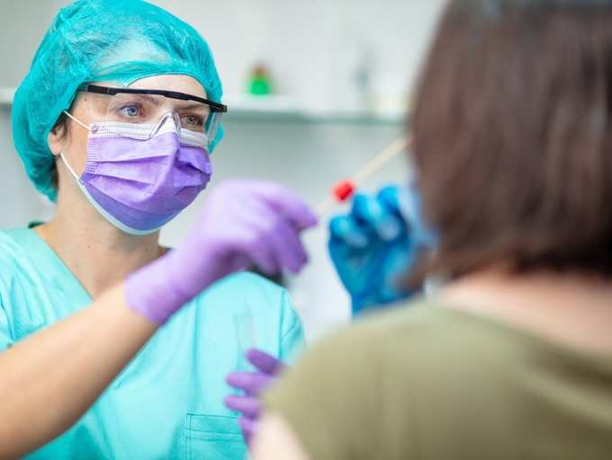
 NO AIR, NO PROBLEM for covid patients:--can we oxygenate blood out side body till lungs get ok

Surviving a COVID-19 ICU stay is just the start. We're ignoring what else  it takes to recover.
Surviving a COVID-19 ICU stay is just ...
nbcnews.com

Extracorporeal Membrane Oxygenation (ECMO)

ECMO stands for extracorporeal membrane oxygenation. The ECMO machine is similar to the heart-lung by-pass machine used in open-heart surgery. It pumps and oxygenates a patient's blood outside the body, allowing the heart and lungs to rest.
.............................................................................................

We Didn’t Evolve for This

A lesson from the animal kingdom on why COVID-19 is so deadly to humans.

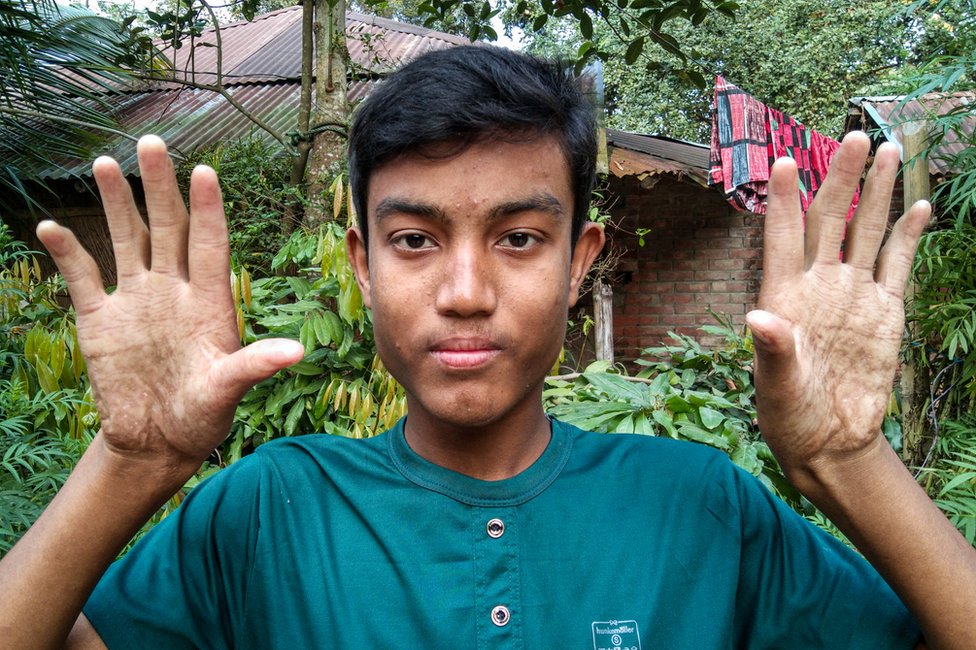
When a Weddell seal, native to Antarctica, plummets 400 meters beneath the ice on one of its hour-long dives, an ensemble of adaptations come together to keep it alive. The seal’s heart rate slows. At this pace, it will burn through its deep reserve of oxygen—provided by extra-large volumes of blood and hemoglobin—more slowly. The seal’s muscles free massive stores of trapped oxygen from another protein, called myoglobin. If oxygen levels become deficient in its tissues, causing hypoxia, cells can use the high levels of the sugar glycogen stored in its heart and brain to begin anaerobic metabolism, creating energy without oxygen. The seal’s extra-large liver also holds its own store of oxygen-rich red blood cells, like a backup scuba tank. And as oxygen levels plummet well below levels that would leave a human diver unconscious, fine control of the veins that oxygenate the seal’s brain cells allow it to swim on unaffected. Together, these systems ensure that the seal survives these intensely hypoxic events again and again, dive after dive, for the many decades of its life.

Geib_BREAKER
NO AIR, NO PROBLEM: Marine mammals, like this seal, comparative anatomist Chris McKnight says, “are this wonderful model, because they live a life that to us just seems like a continuous physiological assault.” Lacking oxygen for long stretches isn’t a problem when you’ve evolved traits to meet that challenge.Steve Rupp

For the past year, humans all over the world have been facing an intensely hypoxic event of their own: COVID-19. The difference? The human body was never built to survive such extreme oxygen restrictions. That fact becomes especially stark when you compare humans to diving marine mammals. 

That is what researchers did in a new paper published in Comparative Biochemistry and Physiology.1 They examined what the extraordinary diving abilities of marine mammals could reveal about what humans face when they contract SARS-CoV-2. The answer they found was, overall, grim: The human body has virtually none of the safeguards that protect a marine mammal when oxygen levels get low.

“Marine mammals have shown us that it takes a lot of coordination, from a lot of tissues, to provide all of that protection in an extreme situation,” said co-author Terrie Williams, a professor at the University of California, Santa Cruz. “And the only thing that we humans can do is make sure that we’re not going to be in a situation where oxygen becomes compromising. Unfortunately, that is exactly what this disease does.”

The human body was never built to survive such extreme oxygen restrictions.

The 
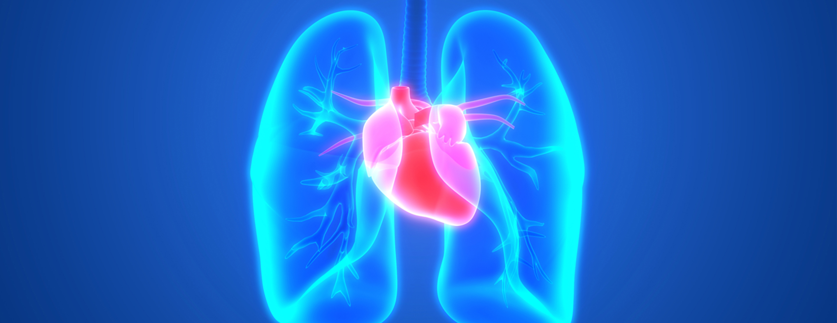
ways that COVID-19 robs human tissues of oxygen is one of its defining characteristics. The virus invades the lining of the lungs, including the alveoli, tiny air sacs that capture oxygen from each breath and quickly pass it into the bloodstream. As the immune system tries to fight off the virus, the lungs and those air sacs become inflamed and fill with fluid, crippling their ability to transport oxygen into the blood. A recent study of patients critically ill from COVID-19 suggests that respiratory failure due to the virus “can be managed similarly to hypoxic respiratory failure” caused by other diseases.2

Many COVID-19 patients also experience abnormal blood clotting, something that scientists and doctors still struggle to explain.3 This clotting can be serious enough to block oxygen from reaching the brain, causing ischemic stroke.4 Brain and heart cells, unlike other kinds of cells, can survive only minutes starved of oxygen before they die.

This cascade of hypoxia reminded Williams—who studies the physiology of both diving mammals and human athletes—of the ways that marine mammals had previously illuminated human health issues. Her research into the hearts of bottlenose dolphins and Weddell seals helped explain a spate of sudden deaths among triathletes as they entered the water at the start of a race: The sudden slap of cold water triggered an instinctive slowing of their hearts, just as they sped up for exercise.5 These findings helped change the format of some races.

On the Origin of Celebrity

I had such fun the other evening. LeBron James, Anne Hathaway, J.K. Rowling, and I had gone ice skating in Central Park. My dear friend Koko the sign-language gorilla was there, ice-dancing with Ryan Gosling, who is always good for...READ MORE

When it comes to COVID-19, however, applying the lessons of deep-diving mammals is not quite as simple. “Marine mammals are this wonderful model, because they live a life that to us just seems like a continuous physiological assault,” says Chris McKnight, a research fellow in comparative anatomy at the Scottish Oceans Institute of St. Andrews University, who was not involved in the paper. “But that comes with its complexity. They’ve had quite a long time to develop those optimal evolutionary traits.”

Even so, some researchers are looking into how those traits evolved to develop treatments. In particular, McKnight pointed to research going on at Duke University’s Cancer Institute. Duke scientists are studying why marine mammals don’t show inflammation in their lungs and other organs when denied oxygen. This protects them from the troubles that human COVID-19 patients have in transporting oxygen to the blood.

Jason Somarelli, an assistant professor at Duke and researcher on this project, explained in an email that his team is studying whether whales may have lost some genes through evolution that allow them to decouple hypoxia and inflammation. If that’s right, it might be possible to one day develop a drug that could artificially turn the same genes off in humans.

“It’s all possible, but part of getting the translation into treatment right is to encourage the biomedical community to pick up the idea that marine mammals may hold keys,” McKnight said. “I wouldn’t imagine there are a huge amount of human biomedical folks whose first stop would be marine mammals as a good place to look.”

To Williams, the lesson from marine mammals is one of caution: They show us just how much evolutionary protection is needed to protect the body from hypoxia, and how few concomitant safeguards humans have. She sees her paper as another way of flagging just how vital it is that people avoid contracting COVID-19 in the first place.


Claudia Geib is a science journalist and editor based on Cape Cod. Her work covers marine and environmental science, wildlife, and how humans connect with the natural world.


References

1. Williams, T.M. & Davis, R.W. Physiological resiliency in diving mammals: Insights on hypoxia protection using the Krogh principle to understand COVID-19 symptoms. Comparative Biochemistry and Physiology 253, 110849 (2020).

2. Hernandez-Romieu, A.C., et al. Timing of intubation and mortality among critically ill coronavirus disease patients: A single-center cohort study. Critical Care Medicine 48, e1045-e1053 (2020).

3. Galiatsatos, P. & Brodsky, R. What does COVID do to your blood? Hopkinsmedicine.org (2020).

4. Szelenberger, R., Saluk-Bijak, J., & Bijak, M. Ischemic stroke among the symptoms caused by the COVID-19 infection. Journal of Clinical Medicine 9, 2688 (2020).

5. Williams, T.M., et al. Exercise at depth alters bradycardia and incidence of cardiac anomalies in deep-diving marine mammals. Nature Communications 6, 6055 (2015).


Extracorporeal Membrane Oxygenation (ECMO)

ECMO stands for extracorporeal membrane oxygenation. The ECMO machine is similar to the heart-lung by-pass machine used in open-heart surgery. It pumps and oxygenates a patient's blood outside the body, allowing the heart and lungs to rest. When you are connected to an ECMO, blood flows through tubing to an artificial lung in the machine that adds oxygen and takes out carbon dioxide; then the blood is warmed to body temperature and pumped back into your body.

There are two types of ECMO. The VA ECMO is connected to both a vein and an artery and is used when there are problems with both the heart and lungs. The VV ECMO is connected to one or more veins, usually near the heart, and is used when the problem is only in the lungs.

USCF is also now using a smaller portable ECMO device that is light enough to be carried by one person and can be transported in an ambulance or helicopter, making it possible to provide ECMO relief in emergency cases.

When is ECMO used:

  • For patients recovering from heart failure, or lung failure or heart surgery.
  • As a bridge option to further treatment, when doctors want to assess the state of other organs such as the kidneys or brain before performing heart or lung surgery.
  • For support during high-risk procedures in the cardiac catheterization lab.
  • As a bridge to a heart assist device, such as left ventricular assist device (LVAD).
  • As a bridge for patients awaiting lung transplant. The ECMO helps keep tissues well oxygenated, which makes the patient a better candidate for transplant.

Procedure

Being placed on ECMO requires a surgical procedure but it is usually done in a patient's room. The patient is sedated and given pain medication and an anti-coagulant to minimize blood clotting. A surgeon, assisted by an operating room team, inserts the ECMO catheters into either an artery or veins. An x-ray is then taken to ensure the tubes are in the right place. Usually a patient on the ECMO pump will also be on a ventilator, which helps the lungs to heal. While on ECMO, the patient will be monitored by specially trained nurses and respiratory therapists, as well as the surgeon and surgical team. Since you will be sedated and have a breathing tube in place, supplemental nutrition will be provided either intravenously or though a nasal-gastric tube. Nutrition is delivered either intravenously or though a nasal-gastric tube

While on ECMO, you may be given certain medications including: heparin to prevent blood clots; antibiotics to prevent infections; sedatives to minimize movement and improve sleep; diuretics to help the kidney get rid of fluids; electrolytes to maintain the proper balance of salts and sugars; and blood products to replace blood loss. Discontinuing ECMO requires a surgical procedure to remove the tubes. Multiple tests are usually done prior to the discontinuation of ECMO therapy to confirm that your heart and lungs are ready. Once the ECMO cannulas are removed, the vessels will need to be repaired. This can be done either at the bedside or in the operating room. The doctor will use small stitches to close the spot where the tubes were placed. You will be asleep and monitored for this process. Even though you are off the ECMO, you may still need to be on a ventilator.

Risks

ECMO does carry risks including:

  • Bleeding, due to the medication that's given to prevent blood from clotting in the tubing.
  • Infection at the sites where the tubes enter the body.
  • Transfusion problems, since a person on ECMO is given blood products.
  • Small clots or air bubbles forming in the tubing.
  • Increased chance of stroke.

UCSF Health medical specialists have reviewed this information. It is for educational purposes only and is not intended to replace the advice of your doctor or other health care provider. We encourage you to discuss any questions or concerns you may have with your provider.

Tuesday, January 19, 2021

coronavirus variants could cause more reinfections, require updated vaccines


Relatives attend a COVID-19 victim’s burial in Manaus, Brazil, on 13 January.

MICHAEL DANTAS/AFP via Getty Images

New coronavirus variants could cause more reinfections, require updated vaccines

Sciences COVID-19 reporting is supported by the Pulitzer Center and the Heising-Simons Foundation.

When the number of COVID-19 cases began to rise again in Manaus, Brazil, in December 2020, Nuno Faria was stunned. The virologist at Imperial College London and associate professor at the University of Oxford had just co-authored a paper in Science estimating that three-quarters of the city’s inhabitants had already been infected with SARS-CoV-2, the pandemic coronavirus—more than enough, it seemed, for herd immunity to develop. The virus should be done with Manaus. Yet hospitals were filling up again. “It was hard to reconcile these two things,” Faria says. He started to hunt for samples he could sequence to find out whether changes in the virus could explain the resurgence.

On 12 January, Faria and his colleagues posted their initial conclusions on the website virological.org. Thirteen of 31 samples collected in mid-December in Manaus turned out to be part of a new viral lineage they called P.1. Much more research is needed, but they say one possibility is that in some people, P.1 eludes the human immune response triggered by the lineage that ravaged the city earlier in 2020.

Emerging variants of the coronavirus have been in the news ever since scientists raised the alarm over B.1.1.7, a SARS-CoV-2 variant that first caught scientists’ attention in England in December and that is more transmissible than previously circulating viruses. But now, they’re also focusing on a potential new threat: variants that could do an end run around the human immune response. Such “immune escapes” could mean more people who have had COVID-19 remain susceptible to reinfection, and that proven vaccines may, at some point, need an update.

At a World Health Organization (WHO) meeting on 12 January, hundreds of researchers discussed the most important scientific questions raised by the wave of new mutations. WHO also convened its COVID-19 Emergency Committee on 14 January to discuss the impact of the new variants and the travel restrictions that many countries are imposing to contain them. The committee called for a global effort to sequence and share more SARS-CoV-2 genomes to help track mutations. It also asked countries to support “global research efforts to better understand critical unknowns about SARS-CoV-2 specific mutations and variants.”

The more transmissible variant, B.1.1.7, is already spreading rapidly in the United Kingdom, Ireland, and Denmark, and probably in many other countries. The U.S. Centers for Disease Control and Prevention released a modeling study on Friday showing the strain could become the predominant variant in the United States in March. But scientists are just as worried about 501Y.V2, a variant detected in South Africa. Some of the mutations it carries, including ones named E484K and K417N, change its surface protein, spike, and have been shown in the lab to reduce how well monoclonal antibodies combat the virus. In a preprint published earlier this month, Jesse Bloom, an evolutionary biologist at the Fred Hutchinson Cancer Research Center, showed that E484K also reduced the potency of convalescent sera from some donors 10-fold—although he is quick to add this does not necessarily mean the mutation would cause people’s immunity to the new strain to drop 10-fold.

P.1 adds to the concerns because it appears to have hit on a similar constellation of mutations and has emerged in a place with a high level of immunity. “Anytime you see the same mutations arising and starting to spread multiple times, in different viral strains across the world, that’s really strong evidence that there’s some evolutionary advantage to those mutations,” Bloom says.

Like B.1.1.7, the variant identified in Manaus is already on the move. Just as Faria was finishing his analysis of the Brazilian genomes, a report was published of a variant detected in travelers arriving in Japan from Brazil—and it turned out to be P.1.

Bad friends

How these new variants are affecting the course of the pandemic is still unclear. In Manaus, for example, P.1 might have nothing to do with the new surge in infections; people’s immunity might simply be waning, says Oxford epidemiologist Oliver Pybus. In a press conference today, WHO’s Mike Ryan cautioned that changes in human behavior are still the major driving force for the resurgence. “It’s too easy to just lay the blame on the variants and say it’s the virus that did it,” he said. “Unfortunately, it’s also what we didn’t do that did it.”

Even if the variant plays a crucial role it might be driving the boost because it is transmitted more easily, like B.1.1.7, not because it can evade the immune response. “Of course it could be a combination of these factors, too,” Pybus says. Similarly, in a recent modeling study, researchers at the London School of Hygiene & Tropical Medicine calculated that South Africa’s 501Y.V2 variant could be 50% more transmissible but no better at evading immunity, or just as transmissible as previous variants but able to evade immunity in one in five people previously infected. “Reality may lie between these extremes,” the authors wrote.

Ester Sabino, a molecular biologist at the University of São Paulo, São Paulo, is launching a study to find reinfections in Manaus that could help decide between these hypotheses for P.1. She is also working to sequence more samples from Manaus from January to follow the variant’s spread. “We don’t have the data yet, but my guess is, it will be at 100% now,” she says. Lab studies investigating the variants are also underway. The United Kingdom today launched a new consortium, G2P-UK (for “genotype to phenotype-UK”), headed by Wendy Barclay of Imperial College London, to study the effects of emerging mutations in SARS-CoV-2. One idea discussed at the 12 January WHO meeting is to set up a biobank that would aid studies by housing virus samples, as well as plasma from vaccine recipients and recovered patients.

Interactions between the new mutations may make it harder to tease out their effects. The variants from the United Kingdom, South Africa, and Manaus all share a mutation named N501Y, for instance, or Nelly, as some researchers call it. But the mutation, which affects the spike protein, also occurs in some variants that do not spread faster, suggesting N501Y does not operate alone, says Kristian Andersen of Scripps Research: “Nelly might be innocent, except maybe when she’s hanging with her bad friends.”

Bloom thinks none of the changes is likely to let the virus escape the immune response entirely. “But I would expect that those viruses have some advantage when a lot of the population has immunity”—which might help explain the surge in Manaus.

Vaccine updates

So far, the virus does not appear to have become resistant to COVID-19 vaccines, says vaccinologist Philip Krause, who chairs a WHO working group on COVID-19 vaccines. “The not-so-good news is that the rapid evolution of these variants suggests that if it is possible for the virus to evolve into a vaccine-resistant phenotype, this may happen sooner than we like,” he adds. That possibility adds to the urgency of putting good surveillance in place to detect such escape variants early on, says biostatistician Natalie Dean of the University of Florida. But it also adds to the urgency of vaccinating people, says Christian Drosten, a virologist at Charité University Hospital in Berlin. “We have to do everything we can now to vaccinate as many people as fast as possible, even if that means running the risk of selecting for some variants,” he says.

If vaccine-resistant SARS-CoV-2 strains emerge, vaccines might need to be updated. Several vaccines could be easily changed to reflect the latest changes, but regulators might balk at authorizing them without seeing updated safety and efficacy data, Krause says. If new variants circulate alongside older strains, multivalent vaccines, effective against several lineages, might even be needed. “To be clear: These are downstream considerations,” Krause says. “The public should not think that this is imminent, and that new vaccines will be needed.” But Ravindra Gupta, a researcher at the University of Cambridge, says manufacturers should start to produce vaccines designed to generate immunity to mutated versions of the spike protein, because they keep cropping up. “It tells us that we should have these mutations in our vaccines, so that you shut off one of the avenues for the virus to go down.”

For now, increased transmissibility is the biggest worry, says virologist Angela Rasmussen of Georgetown University. “I’m puzzled why [that] isn’t a bigger part of the conversation,” she says. The U.S. hospital system, she says, “is at capacity in many places and further increases in transmission can tip us over the edge where the system collapses. Then we’ll start seeing potentially huge increases in mortality.”

doi:10.1126/science.abg6028

Kai Kupferschmidt

Kai is a contributing correspondent for Science magazine based in Berlin, Germany. He is the author of a book about the color blue, published in 2019.

Twitter

===================================

MY COMMENT:-

 VIRUS CAN BE DEFEATED BY DEFENSIVE METHODS AND OFFENSIVE METHODS

NOW WE ARE USING ONLY DEFENSIVE METHODS

[1]
THESE ARE MASK/DISTANCING/QUARANTINE/LOCKDOWN

[2]VACCINES.

BUT EVERYTIME VIRUS MUTATE=THE  VACCINE HAS TO BE CHANGED/UPDATED FOR THE NEW MUTATION
======================================


WHAT WE CAN DO IS ATTACK:-

[1]ATTACK THE CORONA VIRUS USING ANOTHER VIRUSWITH LAB:PRIMED GENES/GENOME TO ATTACK AND KILL CORONA VIRUS
[2]
CHANGE THE GENES/GENOME OF CORONA VIRUS IN LAB: IN SUCH A WAY IT CAN NO LONGER INFECT HUMANS
[3]
CHANGE GENES OF CORONA VIRUS IN LAB:IN SUCH A WAY THAT IT CANNOT MUTATE=CHANGE AS IT IS NOW ;
AND MAKE THE VIRUS DIE OUT FASTER


Friday, January 1, 2021

even RT-PCR tests, considered to be the ‘gold standard’ for testing have been prone to failure rates and wrongful diagnosis.


closecomments

userthumb
loader

Now Reading:

Coronavirus test: Can you get false positives in a COVID-19 test?
Share fbsharetwsharepinshareComments (1)

Coronavirus test: Can you get false positives in a COVID-19 test?

TIMESOFINDIA.COM | Last updated on - Dec 2, 2020, 11:46 ISTShare fbsharetwsharepinshareComments (1)

01/8Can you get false positives in a COVID-19 test?

Preventive testing is one of the ways to fight out the virus, which has infected over 55 million people globally.

While COVID tests under offering right now carry a good accuracy and sensitivity rate, of late, there have been reports of people receiving wrong results, and be wrongly diagnosed with a COVID-19 infection, even when they aren’t.

02/8Can a person be 'wrongly' diagnosed?

Billionaire Elon Musk, grabbed headlines when he talked about the ‘inaccuracy’ of COVID testing after receiving 2 positives and 2 negatives on the same day.

There also have been endless reports of people being wrongly diagnosed with the infection, even while relying on the ‘gold standard’ tests. More recently, an erroneous fault at a UK-based testing lab led to over a 1000 people receiving wrong results on a single day. Loopholes like these have also led many to bat an eye over COVID testing standards, and wonder, how many genuine COVID-19 cases might be really there.

But, the question remains- how can a person get a fake positive result, even when they aren’t really infected?

readmore

03/8What does a false positive mean?

A false positive for COVID-19 means a person who gets a positive COVID diagnosis, despite having no active infection or someone who showcases active antibodies without any infection trace.

While no diagnostic test is 100% accurate, the chances of getting a wrong diagnosis can impact the precedence of a disease. Right now, there are a series of tests available for COVID-19.

The odds of getting false positives are higher with antigen and iG antibody tests. However, even RT-PCR tests, considered to be the ‘gold standard’ for testing have been prone to failure rates and wrongful diagnosis.

We tell you some of the reasons why false positives with COVID-19 can occur, even if rare:

readmore

04/8Different pathology labs can give different results

COVID testing is being done across labs, worldwide. However, COVID-19 test requires a lot more precision and sensitivity to handle than any other diagnostic test, which means that different labs can employ different tools to assess viral load. Misuse of chemicals, diagnostic failures can also be blamed.

This is more common with antigen tests, which dole out results quicker but have a higher chance of throwing up inaccurate results.

05/8Human error to blame?

The pandemic is a sensitive time, and there remains a huge margin of error. COVID testing is being done in huge numbers, and that leaves a big gap for human error, or complacencies to mix up results. It is one of the reasons why a lot of people are receiving a faulty diagnosis in the first place. However, we must remember that this is a rare occurrence, and not always the reason.

06/8Tests can pick up viral ‘debris’

A lot of people who have recovered from COVID-19 continue to test positive for the virus. As strange as that sounds, tests can detect a positive trace even weeks after the contagious period is over, or the symptoms vanish. This is one of the biggest causes of why an RT-PCR test is subject to false positives. Since these tests are highly sensitive and work by amplifying the genetic code of even the tiniest viral fragment, the reverse chain reaction used in the testing process can also pick up viral debris, or the ‘dead’ parts of the virus which could linger in the body and give out a positive result.

Thus, a true recovery from COVID is mapped by the presence of symptoms and not the test results in itself.

readmore

07/8The time you undergo a test matters

Again, while there is no real ‘right’ time to get tested, COVID-19 tests can also throw up wrong results, depending on the timing you get the test done. The chances of getting false negatives exist higher than false positives, but, still, timing is a key factor at play.

08/8False positive vs false negative- What’s more worrisome?

False positive and false negatives can occur with any test. However, considering the pandemic we are facing, false negative results carry worse repercussions, since it puts people who would have been infected with the virus at risk and the ones around them exposed to.

At the same time, experts have also pointed out that even false positives can hinder the role of preventive strategies, making people cloud an air of judgement over the accuracy rates, and in some cases, also cause undue mental harassment, after receiving the diagnosis

......................................................................................................................


False Positive Results in Real-time Reverse Transcription-Polymerase Chain Reaction (rRT-PCR) for SARS-CoV-2?

There are multiple severe acute respiratory syndrome coronavirus 2 (SARS-CoV-2) emergency use authorization (EUA) tests among clinical laboratories and a large amount of cross-assay variation between different assays. Moreover, these assays were rapidly developed, minimally standardized and there is no well recognized external quality assessment program (EQA). As a result, good estimates of the diagnostic sensitivity and specificity are not available. There has been more focus on the diagnostic sensitivity and it is known that sensitivity is poor if the test is performed too early before detectable RNA is shed and that the viral RNA may be detectable for a period of time after active infection although the virus is no longer viable or infective. Therefore, the CDC does not recommend retesting after recovery, but CDC suggests symptomless persons who are immunologically normal are no longer considered infectious about 10 days after symptom onset.

There is less information about diagnostic specificity (false positives). Among others, false positives will depend on the length of the DNA probes, how many and which genes are measured and technical errors. The DNA probes used in the CDC rRT-PCR test kits for SARS-CoV-2 assay are only about 25 bases long which does not meet the FDA recommendation for nucleic acid-based molecular diagnostics for viral disease infections where 100 contiguous bases is desirable (1). Various methods use different genes and different probes that may not be equivalent. There is a 100-fold difference in limit of detection (LoD) between some assays (2). Technical error, especially due to contamination may cause false positives. Seventy-seven professional baseball major league players initially tested positive in one lab but negative elsewhere (3) in what was deemed Lab error. Except that they had multiple sources for testing, they might have been classified as asymptomatic. We don’t know how many other persons were classified in error from this incident.

Originally, PCR was followed by a second step where a separation technique such as a blotting method was used to confirm that the amplified substance was correct. rRT-PCR is usually not followed by a second step. RRT-PCR is usually applied for diagnostic purposes, not for screening. For acute viral infections, after symptoms appear, a rRT-PCR test battery may be performed. In diagnostic testing, symptoms or high-risk behavior cause an increase in prevalence because those with certain symptoms are classified into characterized groups and false positives are few.

Diagnostic applications are usually applied for chronic viral infections such as HCV, HIV and chronic HBV where symptoms or high-risk behavior initiates testing, although there are now screening recommendations for HCV. Still, in all these chronic diseases antibody concentrations are high and serology usually precedes rRT-PCR, so that false positives are rare. At present prevalence, COVID-19 testing is primarily widespread screening without confirmation.

For SARS-CoV-2 rRT-PCR, cycle threshold (Ct) of 24 or less has been shown to be highly predictable for identifying active COVID-19 cases (4), but since LoD of various methods drastically differ it is unclear which methods this applies to. Generally, methods do not amplify more than 40 cycles, but some systems go beyond 40 Ct. It seems likely that short probes in such systems could lead to amplification errors. Although there is no wide spread EQA proficiency programs for SARS-CoV-2, there is one report (5), of EQA in clinical laboratories for other RNA virus. The authors compiled 43 EQAs of rRT-PCR assays, conducted between 2004-2019. Each EQA involved between three and 174 laboratories, which together provided results for 4,113 blind panels containing 10,538 negative samples. 336 of the 10,538 negative samples (3.2%) were reported as positive. The authors defined the lowest percentage of the interquartile range which was 0.8% as a conservative estimate of the false positive rate. In another report, Sin Hang Lee found that 3 of 10 positive proficiency samples in the State of Connecticut were negative containing no SARS-CoV-2 RNA by a confirmatory assay (1). The Foundation for Innovative New Diagnostics (FIND) examined 22 rRT-SARS-CoV-2 diagnostic tests (6) and found diagnostic specificities ranging between 100% and 96% for 100 specimens assayed by each test. Although the great majority showed 100% specificity, given the small number assayed, the lower 95% confidence limit which was 95% for almost all assays would seem to be a better estimate (possible 5% error). Moreover, these were tested under controlled conditions, not at all similar to high output clinical laboratories running thousands of tests.

The Reverend Thomas Bayes (1701-1761) recognized a kind of statistic that predicts the posterior probability from the prior probability. For testing, this means the post test probability can be derived from the pretest probability if the prevalence is known. This sounds complicated but actually, Bayesian statistics are simple compared to classical frequentist statistics since one does not have to apply a null hypothesis, nor interpret p-values or effect-size and the results are obtained from simple mathematics. If, as discussed above (5), a 0.8% false positive rate is correct, at a six percent positive rate that some States claim, then there would be: 100 x 0.06 = 6 positives/100 tests. But if 0.8% are false positives, then only 5.2% are true positives with a positive predictive value (True positives/total positives x 100) of 5.2/6 x 100 = 86.6%. This means about 13.4% are false positive. Notice as the prevalence of disease decreases, the percentage of false positives to total positives increases because the true positive percentage decreases but the percent false positive (in this case 0.8%) stays the same. Thus, the percentage of false positives would be about 26.6% at a three percent positive rate.

The source of the problem is recognized from Bayesian analysis. If the prevalence is low (say a prevalence of 1%) even a very good screening test with 99% diagnostic specificity and 100% sensitivity will produce only 1% false positive results: (diagnostic specificity 1%) = 0.01 x 10,000 tests = 100 false positives/10,000 tests and (0.01% prevalence of disease at 100% sensitivity) = 0.01 x 10,000 = 100 true positive but for a poor positive predictive value of only 50% (100/200 x 100 = 50%). Recognizing this problem, the CDC suggests most testing should be diagnostic: “Considerations for who should get tested: People who have symptoms of COVID-19, people who have had close contact with someone with confirmed COVID-19, people who have been asked or referred to get testing by their healthcare provider, or state health department. Not everyone needs to be tested. (7)”

Because of rightful concern regarding disease transmission from asymptomatic and pre-symptomatic cases, this advice is not being followed. As a result, the great abundance of testing is screening not diagnostic. One way to reduce false positive results is to repeat the test using a test with a different format (different manufacturer). Due to limited testing facilities confirmation is not routinely performed and only a few positives are confirmed by a second rRT-PCR assay. I conclude it is likely that at current active disease prevalence the positive rRT-PCR results of many “asymptomatic” persons are false positives.

There are negative psychological implications of thinking one is infected when one is not and some persons with illness other than COVID-19 who test false positive might be hospitalized with COVID-19 patients and become infected. This may explain why some persons seem to have been infected twice: the first time being a false positive. It seems to me it is important for practicing medical professionals to be aware of these issues so that they can appropriately advise and direct suspect patients for additional testing.

REFERENCES

  1. Lee SH. Testing for SARS-CoV-2 in cellular components by routine nested RT-PCR followed by DNA sequencing International Journal of Geriatrics and Rehabilitation 2020;2:69-96.
  2. FDA. SARS-CoV-2 Reference Panel Comparative Data. Accessed 9/20/2020 doi: 10.1093/cid/ciaa638 https://www.fda.gov/medical-devices/coronavirus-covid-19-and-medical-devices/sars-cov-2-reference-panel-comparative-data.
  3. Maske M. NFL’s 77 positive virus tests were ‘likely false positive results,’ company says. Accessed 9/20/2020 https://www.washingtonpost.com/sports/2020/08/23/nfl-teams-interrupt-practice-schedules-after-positive-coronavirus-tests-new-j
  4. Bullard J, Dust K, Funk D, Strong JE, Alexander D, Garnett L, et al. Predicting infectious SARS-CoV-2 from diagnostic samples. Clin Infect Dis 2020 doi: 10.1093/cid/ciaa638.
  5. Cohen AN, Kessel, B. False positives in reverse transcription PCR testing for SARS-CoV-2. Accessed 9/20/2020 https://doi.org/10.1101/2020.04.26.20080911 (not peer reviewed).
  6. Accessed 10/01/2020 https://www.finddx.org/covid-19/sarscov2-eval-molecular 7. Accessed 10/01/2020 https://www.cdc.gov/coronavirus/2019- ncov/testing/diagnostic-testing.html


  • Tuesday, December 29, 2020

    The family with no fingerprints


    bbc.comwhy BBC and British rulers missed this  from 1750 to 1947 while taxing  and squeezing money from Indians?

    The family with no fingerprints

    At least four generations of Apu Sarker's family have an extremely rare condition leaving them with no fingerprints.

    The family with no fingerprints

    By Mir Sabbir
    BBC Bengalí, Dhaka

    Published

    Apu
    image captionAt least four generations of Apu Sarker's family have an extremely rare condition leaving them with no fingerprints

    Apu Sarker was showing his open palm to me on a video call from his home in Bangladesh. Nothing seemed unusual at first, but as I looked closer I could see the smooth surfaces of his fingertips.

    Apu, who is 22, lives with his family in a village in the northern district of Rajshahi. He was working as a medical assistant until recently. His father and his grandfather were farmers.

    The men in Apu's family appear to share a genetic mutation so rare it is thought to affect only a small handful of families in the world: they have no fingerprints.

    Back in the day of Apu's grandfather, having no fingerprints was no big deal. "I don't think he ever thought of it as a problem," Apu said.

    But over the decades, the tiny grooves that swirl around our fingertips - known properly as dermatoglyphs - have become the world's most collected biometric data. We use them for everything from passing through airports to voting and opening our smartphones.

    An Indian voter gives her fingerprint as she comes to cast her vote at a polling station during India's general electionimage copyrightGetty Images
    image captionA voter in India gives her fingerprint before casting a ballot

    In 2008, when Apu was still a boy, Bangladesh introduced National ID cards for all adults, and the database required a thumbprint. The baffled employees did not know how to issue a card to Apu's father, Amal Sarker. Finally, he received a card with "NO FINGERPRINT" stamped on it.

    In 2010, fingerprints became mandatory for passports and driver's licences. After several attempts, Amal was able to obtain a passport by showing a certificate from a medical board. He has never used it though, partly because he fears the problems he may face at the airport. And though riding a motorbike is essential to his farming work, he has never obtained a driving licence. "I paid the fee, passed the exam, but they did not issue a licence because I couldn't provide fingerprint," he said.

    Amal carries the licence fee payment receipt with him but it doesn't always help him when he gets stopped - he has been fined twice. He explained his condition to both bemused officers, he said, and held up his smooth fingertips for them to see. But neither waived the fine.

    "This is always an embarrassing experience for me," Amal said.

    In 2016, the government made it mandatory to match a fingerprint with the national database in order to purchase a Sim card for a mobile phone.

    "They seemed confused when I went to buy a Sim, their software kept freezing every time I put my finger on the sensor," Apu said, with a wry smile. Apu was denied the purchase, and all the male members of his family now use Sim cards issued in his mother's name.

    Amal
    image captionAmal Sarker's fingertips, missing the unique patterns found on most

    The rare condition likely afflicting the Sarker family is called Adermatoglyphia. It first became widely known in 2007 when Peter Itin, a Swiss dermatologist, was contacted by a woman in the country in her late twenties who was having trouble entering the US. Her face matched the photograph on her passport, but customs officers were not able to record any fingerprints. Because she didn't have any.

    Upon examination, Professor Itin found the woman and eight members of her family had the same strange condition - flat finger pads and a reduced number of sweat glands in the hands. Working with another dermatologist, Eli Sprecher, and graduate student Janna Nousbeck, Professor Itin looked at the DNA of 16 members of the family - seven with fingerprints and nine without.

    "Isolated cases are very rare, and no more than a few families are documented," Prof Itin told the BBC.

    In 2011, the team homed in on one gene, SMARCAD1, which was mutated in the nine printless family members, identifying it as the cause of the rare disease. Virtually nothing was known about the gene at the time. The mutation appeared to cause no other ill-health effects apart from the effects on the hands.

    The mutation they were looking for for those years affected a gene "nobody knew anything about", said Professor Sprecher - hence the years it took to find it. Plus, the mutation affected a very specific part of the gene, he said, "which apparently had no function, in a gene of no function".

    Once discovered, the disease was named Adermatoglyphia, but Prof Itin dubbed it "immigration delay disease", after his first patient's trouble getting into the US, and the name stuck,

    Amal and Apu
    image captionAmal and Apu Sarker. "It is not in my hands, it is something I inherited," Amal said.

    Immigration delay disease can affect generations of a family. Apu Sarker's uncle Gopesh, who lives in Dinajpur, some 350km (217 miles) from Dhaka, had to wait two years to get a passport authorised, he said.

    "I had to travel to Dhaka four or five times in the past two years to convince them I really have the condition," Gopesh said.

    When his office started using a fingerprint attendance system, Gopesh had to convince his superiors to allow him to use the old system - signing an attendance sheet every day.

    A dermatologist in Bangladesh has diagnosed the family's condition as congenital palmoplantar keratoderma, which Prof Itin believes developed into secondary Adermatoglyphia - a version of the disease which can also cause dry skin and reduced sweating on palms and feet - symptoms reported by the Sarkers.

    More testing would be needed to confirm that the family has some form of Adermatoglyphia. Professor Sprecher said his team would be "very glad" to assist the family with genetic testing. The results of those tests might bring the Sarkers some certainty, but no relief from the day to day struggles of navigating the world without fingerprints.

    Apu Sarker's younger brother Anu also inherited the rare gene mutation
    image captionApu Sarker's younger brother Anu also inherited the rare gene mutation

    For the afflicted Sarkers, society seems to be becoming more and more unwieldy, rather than evolving to accommodate their condition. Amal Sarker lived most of his life without too much trouble, he said, but he felt sorry for his children.

    "It is not in my hands, it is something I inherited," he said. "But the way me and my sons are getting in all sorts of problems, for me this is really painful."

    Amal and Apu recently got a new kind of national ID card being issued by the Bangladeshi government, after presenting a medical certificate. The card uses other biometric data too - retina scan and facial recognition.

    But they still can't buy a Sim card or obtain a driver's licence, and obtaining a passport is a long and drawn out process.

    "I am tired of explaining the situation over and over again. I've asked many people for advice, but none of them could give me any definite answer," said Apu. "Someone suggested I go to court. If all options fail, then that's what I might have to do."

    Apu hopes he will be able to get a passport, he said. He would love to travel outside Bangladesh. He just needs to start his application.

    Photographs courtesy of the Sarker family.


    Friday, December 4, 2020

    DrugBank Online | Detailed Drug and Drug Target Information


    DrugBank Online | Detailed Drug and Drug Target Information

    A publicly accessible pharmaceutical knowledge base used by researchers and health professionals globally. Get info on drugs, drug targets, and more.

    Drugs

    Drugs, Targets, Pathways, Indications. Small Molecule ...

    Acetaminophen

    Acetaminophen. Targets (4)Enzymes (15)Carriers (1 ...

    DrugBank Release Version 5.1.7

    Already have an account? Click here to Log In. These DrugBank ...

    Metformin

    Metformin is an antihyperglycemic agent of the biguanide class ...

    About DrugBank

    DrugBank started in 2006 in Dr. David Wishart's lab at the ...

    Morphine

    Morphine, the main alkaloid of opium, was first obtained from ...
    DrugBank's powerful tools and structured pharmaceutical knowledge base enables advances in drug discovery, drug repurposing, precision medicine and ...
    The DrugBank database is a comprehensive, freely accessible, online database containing information on drugs and drug targets. As both a bioinformatics and ...
    Nov 29, 2007 — First released in 2006, DrugBank was designed to serve as a comprehensive, fully searchable in silico drug resource that linked sequence, ...
    by DS Wishart · ‎2008 · ‎Cited by 2231 · ‎Related articles